2010
| News | Registration | Abstracts | Accommodation | Excursions | Deadlines | Organizing committee |
| First circular | Participants | Abstract submission | Travel | Program | Seminar History | Contact us |
| Новости |
| Первый циркуляр |
| Регистрация |
| Оформление тезисов |
| Тезисы |
| Программа |
| Участники |
| Размещение |
| Экскурсии |
| Проезд |
| Важные даты |
| Оргкомитет |
| Обратная связь |
Raman spectroscopy – a versatile tool for the micrometre-range study of minerals and rocks
Nasdala L., Zirbs W.
Institut für Mineralogie und Kristallographie, Universität Wien, Wien, Austria
lutz.nasdala@univie.ac.at
This general lecture aims at imparting analytical knowledge related to the application of the Raman micro-spectroscopy technique (for the basics cf. Orlov et al. 1985; Smith and Carabatos-Nédelec 2001; Nasdala et al. 2004) in studying geological samples. It comprises (i) a brief introduction to the basic physical principles, (ii) some discussion of problems and artefacts related to results of Raman analyses, including those that were published recently in the literature, (iii) an overview of applications in virtually all fields of the Earth sciences, and (iv) a few remarks on the generation and application of Raman-based images of geological objects.
Examples related to the study of alkaline rocks concentrate mainly on kimberlitic diamond specimens. Raman analyses allow one to determine in situ the composition of inclusions, which is an important information in concluding about the chemical composition of the source region (e.g., Nasdala et al. 2003; Brenker et al. 2006). The band up-shift (either of the included phase itself or of the neighbouring host diamond) may be used to quantify remnant compressive strain acting on the inclusion (often also referred to as “remnant pressures”, “overpressures”, or “fossilised pressures”), which in turn may then be used to estimate the formation pressure of the diamond specimen (e.g., Nasdala et al. 2003; Howell et al. 2010). Raman maps can also be used to reveal the internal zoning of diamond crystals, which can for instance be used to study genetic relationships (i.e., the position of inclusions relative to the diamond zoning may help to distinguish protogenetic, syngenetic, and epigenetic inclusions). Finally, first results of a Raman study of natural alpha-irradiation effects in diamond (which are associated with green radio-colouration of this mineral) are presented.
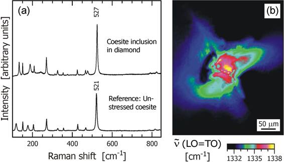
Fig. 1. Two examples for Raman analyses applied in diamond research. (a) Raman spectrum of a coesite inclusion in a large diamond crystal from the Kankan district, Guinea, compared to the reference spectrum of an unstressed coesite (Ries impact crater, Germany). Based on the calibration of Hemley (1987), the observed up-shift of the main coesite band indicates compressive strain of ~2.3 ± 0.2 GPa in the inclusion (cf. Nasdala et al. 2003). (b) Raman map of the same diamond specimen (obtained with E parallel to [001]), visualising a halo of compressive strain in the host diamond around a larnite inclusion. Here, the up-shift of the main diamond band indicates compressive strain up to ~2.6 ± 0.3 GPa (cf. calibration of Grimsditch et al. 1978).
Funding by the European Commission through contract no. MEXC–CT–2005–024878, and the Austrian Science Fund (FWF) through grant P20028–N10, is gratefully acknowledged.
References:
Brenker F.E., Vollmer C., Vincze L., Vekemans B., Szymanski A., Janssens K., Szaloki I., Nasdala L., Joswig W., Kaminsky F. Carbonates from the lower part of transition zone or even the lower mantle. // Earth and Planetary Science Letters. 2006. Vol. 260. P. 1–9.
Grimsditch M.H., Anastassakis E., Cardona M. Effect of uniaxial stress on the zone-center optical phonon of diamond. // Physical Review B. 1978. Vol. 18. P. 901–904.
Hemley R.J. Pressure dependence of Raman spectra of SiO2 polymorphs: a-quartz, coesite and stishovite. // In: Manghnani M.H., Syono Y. (eds.) High-pressure research in mineral physics. Washington D.C.: American Geophysical Union, 1987. P. 347–359.
Howell D., Wood I.G., Dobson D.P., Jones A.P., Nasdala L., Harris J.W. Quantifying strain birefringence halos around inclusions in diamond. // Contributions to Mineralogy and Petrology. 2010. (published online; DOI 10.1007/s00410-010-0503-5).
Nasdala L., Brenker F.E., Glinnemann J., Hofmeister W., Gasparik T., Harris J.W., Stachel T., Reese, I. Spectroscopic 2D-tomography: Residual pressure and strain around mineral inclusions in diamonds. // European Journal of Mineralogy. 2003. Vol. 15. P. 931–935.
Nasdala L., Smith D.C., Kaindl R., Ziemann M. Raman spectroscopy: Analytical perspectives in mineralogical research. // In: Beran A., Libowitzky, E. (eds.) Spectroscopic methods in mineralogy. EMU Notes in Mineralogy. 2004. Vol. 6. P. 281–343.
Nasdala L., Hofmeister W., Harris J.W., Glinnemann J. Growth zoning and strain patterns inside diamond crystals as revealed by Raman maps. // American Mineralogist. 2005. Vol. 90. P. 745–748.
Orlov R.Yu., Uspenskaya M.E., Guseva E.V. Appplication of Raman spectroscopy in mineralogy (in Russian). Moscow: Izd. MGU, 1985. 112 p.
Smith D.C., Carabatos-Nédelec C. Raman spectroscopy applied to crystals: Phenomena and principles, concepts and conventions. // In: Lewis I.R., Edwards H.G.M. (eds.) Handbook of Raman spectroscopy. From the research laboratory to the process line. Practical spectroscopy series Vol. 28. 2001. New York, Basel: CRC Press. P. 349-422.